Many surfaces found in nature possess sophisticated topographical structures which provide them with exceptional properties. Nano-structured surface of lotus leafs efficiently repel water, microscopic brushes of gecko fingers drastically increase the contact with surfaces, topographical structures provides for enhanced anti-reflection surface properties in insects. Self-cleaning ability of bio-surfaces enables them to prevent contamination by particles such as dust, spores, and bacteria.

While investigating the initial adhesion behaviour of Pseudomonas aeruginosa ATCC 9027T and Staphylococcus aureus CIP 65.8T bacterial cells on the surface of cicada Psaltoda claripennis cicada wings, we found another striking phenomenon, namely the wing surfaces resisted colonisation by S. aureus and were lethal for P. aeruginosa bacterial cells (Ivanova 2012).

This is the first reported example of a naturally existing surface with a physical structure that exhibits such effective bactericidal properties. Cicada wing nanopillars are extremely effective at killing Pseudomonas aeruginosa cells; the wing surface was able to kill individual cells within approximately 3 min. This bactericidal ability of the wing surface is primarily a physico-mechanical effect, as it is retained when the surface chemistry is substantially altered (Ivanova 2012, Pogodin 2013).
[/styled_box]Usually when thinking about anti-bacterial surfaces, one considers the surfaces repelling bacteria and preventing the attachment to the surface and consequent formation of a biofilm (Antibiofouling, commonly referred to simply as antifouling, is the property possessed by some materials which prevent the adhesion and subsequent build-up of biological matter on their surfaces). Our investigations into the interaction of Pseudomonas aeruginosa cells with the cicada wings revealed that the wing surface was actually not particularly effective at repelling the bacteria, as many cells were able to adhere to the wing surface, but in contrast, attract bacterial cell wall and break it.
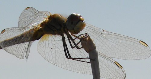
The structure of Psaltoda claripennis cicadae wings are characterized by a series of longitudinal veins, cross veins and the areas enclosed by these regions, known as cells (Ivanova 2012).

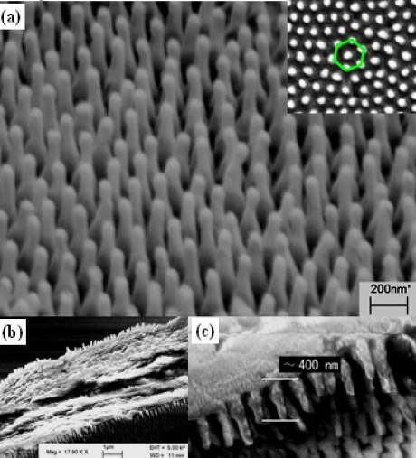
The ventral and dorsal sides of both the fore and hind wings are covered with a periodic topography consisting of hexagonal arrays of spherically-capped, conical, nanoscale pillars (Ivanova 2012):
The height, spacing and diameter of the nanopillars varies between species, however for Psaltoda claripennis they are 200 nm tall, 100 nm in diameter at the base, 60 nm in diameter at the cap, and spaced 170 nm apart from centre to centre. Spatial variations in the wettability of the wing surface was assessed by measuring water droplet contact angles in a grid pattern, before generating a map, coloured according to contact angle. The nanostructured wing surface is hydrophobic; the average water contact angles measured on the wing was 158.8° (Ivanova 2012, Pogodin 2013).

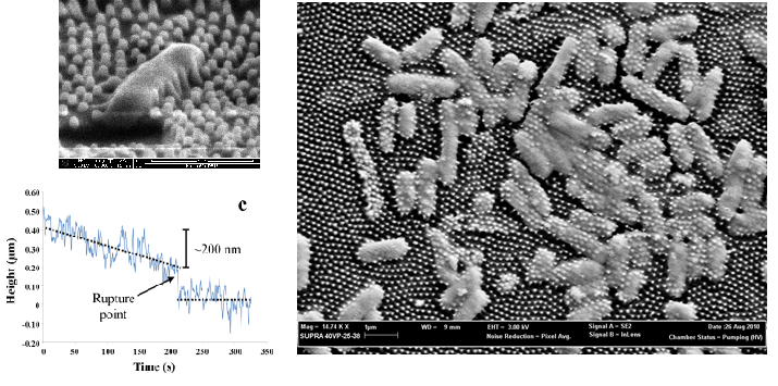
The bacterial cells’ morphology was significantly altered when in contact with the cicada wing surface. The pattern induces rupture of P. aeruginosa bacterial cells (Ivanova 2012, Pogodin 2013).
On scanning electron micrographs the cells can be clearly seen to be penetrated by the nanopillar structures of the wing surface. The wing nano-pillars appeared to penetrate all of the cells that had attached to the surface. Cellular components appeared to be spreading outwards beneath the cell between the nanopillars. Subsequent viability analysis of these cells revealed that these penetrated cells were in fact dead (Ivanova 2012, Pogodin 2013).
While many superhydrophobic surfaces do inhibit the attachment of bacterial cells, we have also shown that there is no direct relationship between superhydrophobicity and antibiofouling. The cicada wings do not actually possess any appreciable antibiofouling ability with respect to Pseudomonas aeruginosa; however the net result is somewhat similar in that the bacteria are prevented from proliferating on the surface.
References:
1) Elena P. Ivanova, Jafar Hasan, Hayden K. Webb, Vi Khanh Truong, Gregory S. Watson, Jolanta A. Watson, Vladimir A. Baulin, Sergey Pogodin, James Y. Wang, Mark J. Tobin, Christian Löbbe, Russell J. Crawford. Natural bactericidal surfaces: mechanical rupture of Pseudomonas aeruginosa cells by cicada wings // Small. 2012. V. 8(16). P. 2489–94. Doi:10.1002/smll.201200528.
2) Jafar Hasan, Hayden K. Webb, Vi Khanh Truong, Sergey Pogodin, Vladimir A. Baulin, Gregory S. Watson, Jolanta A. Watson, Russell J. Crawford, Elena P. Ivanova. Selective bactericidal activity of nanopatterned superhydrophobic cicada Psaltoda claripennis wing surfaces // Applied microbiology and biotechnology. 2012. Doi:10.1007/s00253-012-4628-5.
3) Sergey Pogodin, Jafar Hasan, Vladimir A. Baulin, Hayden K. Webb, Vi Khanh Truong, The Hong Phong Nguyen, Veselin Boshkovikj, Christopher J. Fluke, Gregory S. Watson, Jolanta A. Watson, Russell J. Crawford and Elena P. Ivanova. Biophysical model of bacterial cell interactions with nanopatterned cicada wing surfaces // Biophysical journal. 2013. V. 104(4). P. 835–40. Doi:10.1016/j.bpj.2012.12.046.
Network profiles:
name=”Elena Ivanova”
photo=”https://softmat.net/wp-content/uploads/2012/05/ivanova.jpg”
url=”https://softmat.net/t2t_products/ivanova/”
] [people_short
name=”Vladimir Baulin”
photo=”https://softmat.net/wp-content/uploads/2012/05/baulin1.jpg”
url=”https://softmat.net/t2t_products/baulin/”
]
